Scroll to:
Crystalline structure of polyacrylonitrile- and viscose-based carbon fibers following high-temperature treatment in the range of 1500–2800 °C
https://doi.org/10.17073/1997-308X-2025-1-30-39
Abstract
The crystalline structure of carbon fibers (CF) based on polyacrylonitrile (PAN) and viscose precursors, treated in the temperature range of 1500 to 2800 °C, was studied using X-ray diffraction analysis and Raman spectroscopy. The objective of the study was to obtain data on the structure of low-modulus viscose-based fibers, which are widely used as fillers in composite materials, and to compare the characteristics of CF derived from different precursors. An empirical dependence of the intensity ratio of the D and G lines (ID /IG ) of the Raman spectra on the treatment temperature was established for carbon fibers based on viscose and PAN. The crystallite sizes La and Lc of both types of CF obtained at different treatment temperatures were evaluated. It was revealed that as the treatment temperature increases, the crystallite sizes La and Lc grow, while the interlayer spacing d002 decreases, indicating an increase in the degree of graphitization. It was found that viscose-based carbon fibers exhibit a less ordered crystalline structure compared to PAN fibers processed under the same conditions. Additionally, the true density and elastic modulus of viscose-based CF were investigated, showing lower values than those of PAN fibers treated at the same temperature. These differences in the properties and structure of CF are attributed to the microtextured nature of viscose fibers. However, during treatment at 2800 °C, CF undergo partial graphitization, which significantly reduces structural differences between fibers of both types. Nevertheless, despite the similarity in crystalline structure, viscose-based CF, even after high-temperature treatment, does not become analogous to PAN-based fibers.
For citations:
Kleusov B.S., Samoilov V.M., Elchaninova V.A., Budushin D.A., Litovchenko E.M., Poplavskaya A.S., Vorontsov V.A. Crystalline structure of polyacrylonitrile- and viscose-based carbon fibers following high-temperature treatment in the range of 1500–2800 °C. Powder Metallurgy аnd Functional Coatings (Izvestiya Vuzov. Poroshkovaya Metallurgiya i Funktsional'nye Pokrytiya). 2025;19(1):30-39. https://doi.org/10.17073/1997-308X-2025-1-30-39
Introduction
The development of carbon fiber-reinforced plastics has led to the production of a wide range of carbon fibers (CF) [1–6]. The existing classification divides all CF into several types based on their modulus: low-modulus (30–100 GPa), intermediate-modulus and high-strength (200–350 GPa), high-modulus (350–500 GPa), and ultra-high-modulus (500–1000 GPa) [6–11]. Another critical factor in classifying fibers is the precursor type, which determines the crystalline structure of CF and, ultimately, its final properties [6–11]. Currently, nearly all commercially produced CF are derived from three main precursors: polyacrylonitrile (PAN), isotropic and mesophase pitches, and viscose [6–11].
The crystalline structure of PAN- and mesophase pitch-based CF has been extensively studied using X-ray diffraction, often in combination with Raman spectroscopy and electron microscopy [12–17]. However, the structure of viscose-based fibers remains underexplored. Existing data in early literature [18; 19] pertain to the technology for producing intermediate- and high-modulus viscose-based CF, developed over 50 years ago in the United States. Studies on the crystalline structure of low-modulus (30–100 GPa) viscose-based CF are extremely limited [20–22], despite their widespread use as fillers in composite materials for various applications.
The aim of this study was to investigate the crystalline structure of viscose-based carbon fibers and its changes during high-temperature treatment, with a comparative analysis of similar data for PAN-based CF.
Материалы и методы исследования
For this study, semi-finished products of TGN-brand viscose-based CFs and UKN-type PAN-based CFs, both manufactured in the Russian Federation, were used. The samples were obtained by additional heat treatment (HT) of CF bundles in a laboratory Tammann furnace under argon atmosphere in a free state (without tension). The heating rate was 300 °C/h, and the dwell time at the target temperature was 20 min. The processing temperature was monitored using a pyrometer.
The true density of the obtained CF samples was measured using the gradient tube method in accordance with GOST R ISO 10119–2012. The average filament diameter, tensile strength, and dynamic elastic modulus of single filaments were measured according to ASTM D4018–11. The physical and mechanical properties of CF were determined as averages from 25 measurements of tensile strength and elastic modulus, following GOST 6943.5–79 and GOST 28008–88.
Raman spectra of CF subjected to various HT temperatures (tHT ) were recorded from the lateral surface of filaments in the broad spectral range of 700–3000 cm\(^{−1}\) using a confocal Raman microspectrometer Via Reflex (Renishaw, UK) equipped with an optical microscope and a cooled CCD detector. The laser spot size at 100× magnification was 0.5 µm. The excitation source was a diode-pumped solid-state Nd:YAG laser with a wavelength of 532 nm and a power of 1 mW.
In the first-order spectrum (1000–2000 cm\(^{−1}\)), carbon materials, including CF, typically exhibit two characteristic bands [30; 31; 34]. One is the band at ν = 1580 cm\(^{−1}\), allowed by Raman scattering and corresponding to the ideal graphitic vibrational mode with E2g symmetry, often referred to as the G mode [23–27]. It is associated with in-plane vibrations of carbon atoms in graphene layers and relates to carbon atoms in an sp2-hybridized state. The other band, at ν = 1360 cm\(^{−1}\), is due to disordered carbon atoms, corresponds to lattice vibrations with A1g symmetry, and is called the D mode [23–27]. This mode is linked to carbon atoms in sp2- and sp3 hybridization states, typically localized at defects or the edges of graphene layers [23–27]. The D band is absent in monocrystalline graphite, and its increasing intensity is considered indicative of a higher content of disordered or peripheral carbon [23–27]. According to numerous studies, for crystallite sizes up to 2 nm, the ratio of the integrated intensities of these bands (ID /IG ) depends on the defect concentration and follows the Ferrari equation [28; 30–32]. For crystallite sizes larger than 2 nm, the ID /IG ratio is determined by the average distance between defects. In graphitizing carbon materials, it can characterize the average crystallite size (La ) using the Tuinstra-Koenig relation [29–31]. For the studied CF, La was calculated using the following equation:
| \[\frac{{{I_D}}}{{{I_G}}} = \frac{{C(\lambda )}}{{{L_a}}},\] | (1) |
where C(λ) is a constant dependent on the laser wavelength. Thus, C(λ = 532 nm) is approximately equal to 4.4 nm [23; 24; 27].
The interpretation of the secondary 2D band (ν = 2700 cm\(^{−1}\)) is more complex. This band appears at a sufficiently high degree of crystalline structure perfection and typically consists of several components [24; 27]. However, for the purposes of this study, only the tHT (heat-treatment temperature) at which the 2D band appears was recorded.
X-ray phase analysis was conducted using a D8 Advance diffractometer (Bruker, Germany). A copper X-ray tube with a maximum power of 2200 W and CuKα radiation (λ = 0.15418 nm) was used as the X-ray source, in Bragg–Brentano geometry (reflection mode). X-ray diffraction patterns were recorded over the angular range of 2θ = 10÷90°, with a scanning speed of 2°/min and a step size of 0.02°. The fibers were placed on a low-background silicon holder, evenly distributed over its surface. Before each measurement, the tube and detector were initialized. The diffraction patterns were processed using the specialized TOPAS software. The absolute error in measuring the angular positions of diffraction peaks did not exceed ±0.026° [33]. The interplanar spacing (d002) was calculated based on the center of gravity of the (002) peak using the Wulff–Bragg equation:
| \[{d_{002}} = \frac{\lambda }{{2\sin {\theta _{002}}}},\] | (2) |
where λ is the wavelength of the X-ray radiation and θ002 is the diffraction angle determined from the center of gravity of the (002) reflection.
The crystallite sizes were calculated using the Scherrer formula:
| \[{L_c} = \frac{{k\lambda }}{{\beta \cos {\theta _{002}}}},\] | (3) |
where β is the full width at half maximum (FWHM) of the (002) reflection, and k = 0.89 [32; 33].
Results and discussion
Fig. 1 presents photographs of PAN- and viscose-based CF filaments treated at processing temperatures tHT = 1200 and 2800 °С. It is evident that the microstructure of the fracture surface and the lateral surface of the filaments of the studied CF at tHT = 1200 °С show minimal differences. However, the fracture surface photographs of the CF after heat treatment at 2800 °С exhibit pronounced differences.
Fig. 1. Photographs of viscose-based CF (а, b) and PAN-based CF filaments (c, d) at |
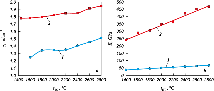
The dependencies of the true density of CF filaments (γ, g/cm3) and the dynamic elastic modulus (E, GPa) on the processing temperature of the studied fibers are shown in Fig. 2. It can be seen that viscose-based fibers exhibit lower values of γ and Е compared to PAN-based CF across the entire range of tHT . Notably, the elastic modulus of viscose-based fibers is 4–5 times lower than that of PAN-based fibers throughout the temperature range.
Fig. 2. Dependence of true density (a) and dynamic elastic modulus (b) |
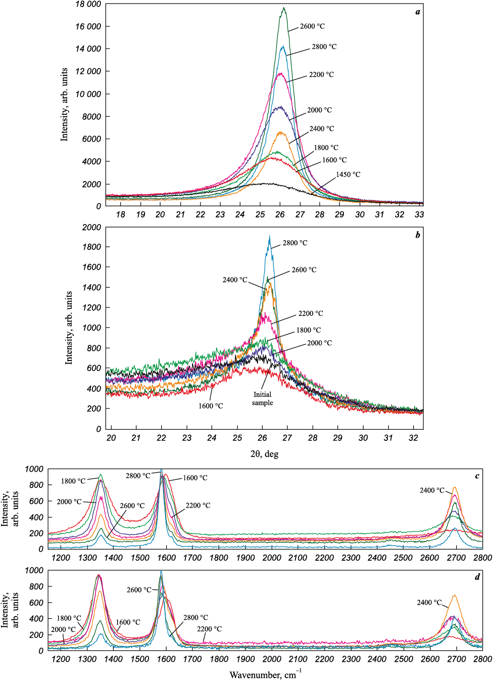
Fig. 3 shows the X-ray diffraction patterns and Raman spectra of the studied CF with varying processing temperatures, while Fig. 4 illustrates the dependence of their crystalline structure parameters on tHT .
Fig. 3. X-ray diffraction patterns of CFs (а, b) based on PAN (a), and viscose (b), |
It is evident that the increase in intensity and narrowing of the (002) diffraction line indicates an improvement in the crystalline structure with increasing tHT for both viscose- and PAN-based CF (Fig. 3, a, b). The asymmetry of the diffraction peak can be effectively described by multiple structural components [34–35]; however, this study provides averaged data for one of these components.
In the Raman spectra of the studied CF (Fig. 3, c, d), the D and G bands become narrower with increasing tHT , and the relative intensity of the D peak decreases. After heat treatment at t ~ 1800 °C, the 2D peak appears, and its intensity relative to the G peak increases with rising processing temperature.
Fig. 4. Dependence of crystalline structure parameters on the processing temperature |
However, after heat treatment at 2800 °C, the differences in the crystalline structure parameters of viscose- and PAN-based CF become insignificant or disappear entirely (see Fig. 3), except for the crystallite size La (see Fig. 4).
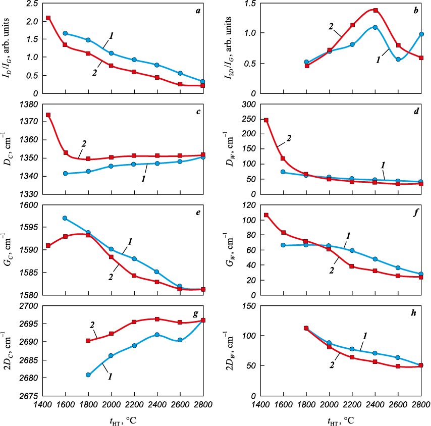
Fig. 5 shows the dependencies of Raman spectroscopy parameters for viscose-based CFs (1) and PAN-based CFs (2) on the processing temperature.
Fig. 5. Dependence of Raman spectroscopy parameters for viscose-based CFs (1) |
It is evident that the positions and widths of the D and G bands systematically change with increasing tHT . In accordance with the results of previous studies, the dependence of the ID /IG parameter was previously used by us to evaluate the effective processing temperature of PAN-based CFs [36].
Using a similar approach, empirical expressions for determining the effective processing temperature (teff , °С) of PAN-based (4) and viscose-based (5) CFs were derived based on the obtained dependencies of the ID /IG parameter on tHT (see Fig. 5, а):
| \[{t_{{\rm{eff}}}} = 2089 - \left( {901\ln \frac{{{I_D}}}{{{I_G}}}} \right),\] | (4) |
| \[{t_{{\rm{eff}}}} = 1815 - \left( {641\ln \frac{{{I_D}}}{{{I_G}}}} \right).\] | (5) |
Conclusion
The results of the study indicate that viscose-based carbon fibers (CFs) exhibit a significantly lower degree of crystalline structure perfection compared to PAN-based CFs across almost the entire range of heat treatment temperatures. However, high-temperature treatment at 2800 °C largely mitigates these differences, suggesting partial graphitization of viscose-based CFs. Nevertheless, as evidenced by the full set of obtained data, despite the similarity in most crystalline structure parameters, viscose-based CFs do not become equivalent to PAN-based CFs even after high-temperature treatment. The elastic modulus of viscose-based CFs does not exceed 100 GPa, which is over four times lower the elastic modulus of PAN-based CFs subjected to identical treatment conditions. The true density of viscose-based CFs remains significantly lower compared to PAN-based CFs (see Fig. 2, a), indicating the distinctive nature of their porosity.
These differences, in our view, can be attributed to the inherently low degree of microtexturing in viscose-based CFs compared to PAN-based CFs and, to an even greater extent, to mesophase pitch-based CFs [19; 37]. The closest counterpart to low-modulus viscose-based CFs are fibers produced from isotropic pitches [10], which similarly exhibit reduced true density and microtexturing.
Based on previous studies [7; 22], which found no significant differences in the properties of the raw viscose fibers used for CF production, it can be inferred that the low elastic modulus of the investigated viscose-based CFs is primarily due to the absence of intensive orientational stretching during the graphitization process.
References
1. Gupta M.K., Singhal V., Rajput N.S. Applications and challenges of carbon-fibres reinforced composites: A review. Evengreen Joint Journal of Novel Carbon Resource Sciences & Green Asia Strategy. 2022;9(3):682–693. https://doi.org/10.5109/4843099
2. Ince J.C., Peerzada M., Mathews L.D., Pai A.R., Al-qatatsheh A., Abbasi S., Yin Y., Hameed N., Duffy A.R., Lau A.K., Salim N.V. Overview of emerging hybrid and composite materials for space applications. Advanced Composites and Hybrid Materials. 2023;6(4):130. https://doi.org/10.1007/s42114-023-00678-5
3. Zhao J. Carbon fiber applications in modern rockets. Preprint. June 2023. https://doi.org/10.13140/RG.2.2.12957.49126
4. Olofin I., Liu R. The application of carbon fibre reinforced polymer (CFRP) cables in civil engineering structures. International Journal of Civil Engineering. 2015;2(7):1–5. https://doi.org/10.14445/23488352/IJCE-V2I7P101
5. Ozkan D., Gok M.S., Karaoglanli A.C. Carbon fiber reinforced polymer (CFRP) composite materials, their characteristic properties, industrial application areas and their machinability. In: Öchsner A., Altenbach H. (eds). Engineering Design Applications III. Advanced Structured Materials, vol. 124. Springer, Cham. 2020. Р. 235–253. https://doi.org/10.1007/978-3-030-39062-4_20
6. Morgan P. Carbon fibers and their composites. London: Taylor & Francis Group, 2005. 1166 p. https://doi.org/10.1201/9781420028744
7. Park S.-J., Heo G.-Y. Precursors and manufacturing of carbon fibers. In: Carbon Fibers. Springer Series in Materials Science, vol. 210. Springer, Dordrecht. 2014. P. 31–66. https://doi.org/10.1007/978-94-017-9478-7_2
8. Emmerich F.G. Young’s modulus, thermal conductivity, electrical resistivity and coefficient of thermal expansion of mesophase pitch-basedcarbonfibers. Carbon. 2014;79:274–293. https://doi.org/10.1016/j.carbon.2014.07.068
9. Frank E., Steudle L.M., Ingildeev D., Spörl J.M., Buchmeiser M.R. Carbon fibers: Precursor systems, processing, structure, and properties. Angewandte Chemie International Edition. 2014;53(21):5262–5298. https://doi.org/10.1002/anie.201306129
10. Newcomb B.A. Processing, structure, and properties of carbon. Composites Part A: Applied Science and Manufacturing. 2016;91:262–282. https://doi.org/10.1016/j.compositesa.2016.10.018
11. Mirdehghan S.A. Fibrous polymeric composites. In: Engineered polymeric fibrous materials. The Textile Institute. Book Series. Woodhead Publishing, 2021. Р. 1–58. https://doi.org/10.1016/B978-0-12-824381-7.00012-3
12. Qiu L., Zheng X.H., Zhu J., Su G.P., Tang D.W. The effect of grain size on the lattice thermal conductivity of an individual polyacrylonitrile-based carbon fiber. Carbon. 2013;51:265–273. https://doi.org/10.1016/j.carbon.2012.08.052
13. Sun Z., Lu Y., Wang R., Yang C. Analysis of carbon fiber structure based on dynamic laser Raman spectroscopy. Journal of Applied Polymer Science. 2020;138(16):50247. https://doi.org/10.1002/app.50247
14. Li D., Wang H., Wang X. Effect of microstructure on the modulus of PAN-based carbon fibers during high temperature treatment and hot stretching graphitization. Journal of Materials Science. 2007;42(12):4642–4649. https://doi.org/10.1007/s10853-006-0519-4
15. Zhou G., Liu Y., He L., Guo Q., Ye H. Microstructure difference between core and skin of T700 carbon fibers in heat-treated carbon/carbon composites. Carbon. 2011;49(9):2883–2892. https://doi.org/10.1016/j.carbon.2011.02.025
16. Wu G.-P., Li D.-H., Yang Y., Lu C.-X., Zhang S.-C., Li X.-T., Feng Z.-H., Li Z.-H. Carbon layer structures and thermal conductivity of graphitized carbon fibers. Journal of Materials Science. 2011;47(6):2882–2890. https://doi.org/10.1007/s10853-011-6118-z
17. Qin X., Lu Y., Xiao H., Wen Y., Yu T. A comparison of the effect of graphitization on microstructures and properties of polyacrylonitrile and mesophase pitch-based carbon fibers. Carbon. 2012;50(12):4459–4469. https://doi.org/10.1016/j.carbon.2012.05.024
18. Sarian S., Strong S.L. Mechanical properties of stress-graphitised carbon fibers: Thermally induced relaxation and recovery. Fibre Science and Technology. 1971;4(1):67–79. https://doi.org/10.1016/0015-0568(71)90012-1
19. Diefendorf R.J., Tokarsky E. High-performance carbon fibers. Polymer Engineering and Science. 1975;15(3): 150–159. https://doi.org/10.1002/pen.760150306
20. Spörl J. M., Ota A., Son S., Massonne K., Hermanutz F., Buchmeiser M.R. Carbon fibers prepared from ionic liquid-derived cellulose precursors. Materials Today Communications. 2016;7:1–10. https://doi.org/10.1016/j.mtcomm.2016.02.002
21. Bengtsson A., Bengtsson J., Sedin M., Sjöholm E. Carbon fibres from lignin-cellulose precursors: Effect of stabilisation conditions. ACS Sustainable Chemistry & Engineering. 2019;7(9):8440–8448. https://doi.org/10.1021/acssuschemeng.9b00108
22. Dumanli A.G., Windle A.H. Carbon fibres from cellulosic precursors: A review. Journal of Materials Science. 2012;47(10):4236–4250. https://doi.org/10.1007/s10853-011-6081-8
23. Tuinstra F., Koenig J.L. Raman spectrum of graphite. The Journal of Chemical Physics. 1970;53(3):1126–1130. https://doi.org/10.1063/1.1674108
24. Cançado L.G., Takai K., Enoki T., Endo M., Kim Y.A., Mizusaki H., Jorio A., Coelho L.N., Magalhães Paniago R., Pimenta M.A. General equation for the determination of the crystallite size La of nanographite by Raman spectroscopy. Applied Physics Letters. 2006;88(16):3106–3109. https://doi.org/10.1063/1.2196057
25. Reich S., Thomsen C. Raman spectroscopy of graphite. Philosophical Transactions of the Royal Society A: Mathematical, Physical and Engineering Sciences. 2004; 362(1824):2271–2288. https://doi.org/10.1098/rsta.2004.1454
26. Ferrari A.C., Robertson J. Interpretation of Raman spectra of disordered and amorphous carbon. Physical Review B. 2000;61(20):14095–14107. https://doi.org/10.1103/physrevb.61.14095
27. Cancado L.G., Jorio A., Martins Ferreira E.H., Stavale F., Achete C.A., Capaz R.B., Moutinho M.V.O., Lombardo A., Kulmala T.S., Ferrari A.C. Quantifying defects in graphene via Raman spectroscopy at different excitation energies. Nano Letters. 2011;11(8):3190–3196. https://doi.org/10.1021/nl201432g
28. Ferrari A.C., Basko D.M. Raman spectroscopy as a versatile tool for studying the properties of graphene. Nature Nanotechnology. 2013;8(4):235–246. https://doi.org/10.1038/nnano.2013.46
29. Zickler Gerald A., Smarsly B., Gierlinger N., Peterlik H., Paris O.A reconsideration of the relationship between the crystallite size La of carbons determined by X-ray diffraction and Raman spectroscopy. Carbon. 2006;44(15): 3239−3246. https://doi.org/10.1016/j.carbon.2006.06.029
30. Okuda H., Young R. J., Wolverson D., Tanaka F., Yamamoto G., Okabe T. Investigating nanostructures in carbon fibres using Raman spectroscopy. Carbon. 2018; 130:178–184. https://doi.org/10.1016/j.carbon.2017.12.108
31. Samoilov V.M., Samsonova V.B., Nakhodnova A.V., Verbets D.B., Gareev A.R., Bubnenkov I.A., Steparyova N.N., Shvetsov A.A., Bardin N.G. Raman spectroscopy and crystalline structure of polyacrylonitrile-based carbon fibres. Advanced Materials & Technologies. 2019;3(15):8–15. https://doi.org/10.17277/amt.2019.03.pp.008-015
32. Pacault A. Chemistry and physics of carbon. Ed. L. Walker. Vol. 7. N.Y.: Marcel Dekker, 1971. 403 p.
33. Cheblakova E.G. , Kleusov B.S., Sapozhnikov V.I. , Gorina V.A., Malinina Yu.A., Gareev A.R. Investigation of the properties of high-strength fibers by methods of physico-chemical analysis. Powder Metallurgy аnd Functional Coatings. 2023;17(4):34–40. https://doi.org/10.17073/1997-308X-2023-4-34-40
34. Tyumentsev V.A., Fazlitdinova A.G., Podkopaev S.A., Churikov V.V. Fine structure of polyacrylonitrile and carbon fibers. Izvestiya vuzov. Khimiya i khimicheskaya tekhnologiya. 2013;56(7):83–87. (In Russ.).
35. Tyumentsev V.A., Fazlitdinova A.G. Study of the structure of fibrous carbon materials by X-ray diffractometry. Zavodskaya laboratoriya. Diagnostika materialov. 2019; 85(11):31–36. (In Russ.). https://doi.org/10.26896/1028-6861-2019-85-11-31-36
36. Samoilov V.M., Nakhodnova A.V., Osmova M.A., Verbets D.B., Bubnenkov A.N., Steparyova N.N., Gareev A.R., Fateeva M.A., Shilo D.V., Ovsyannikov N.E. Effective heat treatment temperature of carbon materials in high temperature furnaces: determination by the parameters of raman spectroscopy of witness samples. Inorganic Materials: Applied Research. 2021;12(5):1416–1427. https://doi.org/10.1134/S2075113321050348
37. Northolt M.G., Veldhuizen L.H., Jansen H. Tensile deformation of carbon fibers and the relationship with the modulus for shear between the basal planes. Carbon. 1991;29(8):1267–1279. https://doi.org/10.1016/0008-6223(91)90046-l
About the Authors
B. S. KleusovRussian Federation
Boris S. Kleusov – Senior Researcher, Testing Center
1 Bld, 2 Electrodnaya Str., Moscow 111524, Russia
V. M. Samoilov
Russian Federation
Vladimir M. Samoilov – Chief Researcher
1 Bld, 2 Electrodnaya Str., Moscow 111524, Russia
V. A. Elchaninova
Russian Federation
Victoria A. Elchavinova – Researcher
1 Bld, 2 Electrodnaya Str., Moscow 111524, Russia
D. A. Budushin
Russian Federation
Dmitry A. Budushin – Intern Researcher
1 Bld, 2 Electrodnaya Str., Moscow 111524, Russia
E. M. Litovchenko
Russian Federation
Egor M. Litovchenko – Student
1 Bld, 20 Geroev Panfilovtsev Str., Moscow 125480, Russia
A. S. Poplavskaya
Russian Federation
Anna S. Poplavskaya – Engineer
1 Bld, 2 Electrodnaya Str., Moscow 111524, Russia
V. A. Vorontsov
Russian Federation
Vladimir A. Vorontsov – Department Head
1 Bld, 2 Electrodnaya Str., Moscow 111524, Russia
Review
For citations:
Kleusov B.S., Samoilov V.M., Elchaninova V.A., Budushin D.A., Litovchenko E.M., Poplavskaya A.S., Vorontsov V.A. Crystalline structure of polyacrylonitrile- and viscose-based carbon fibers following high-temperature treatment in the range of 1500–2800 °C. Powder Metallurgy аnd Functional Coatings (Izvestiya Vuzov. Poroshkovaya Metallurgiya i Funktsional'nye Pokrytiya). 2025;19(1):30-39. https://doi.org/10.17073/1997-308X-2025-1-30-39


